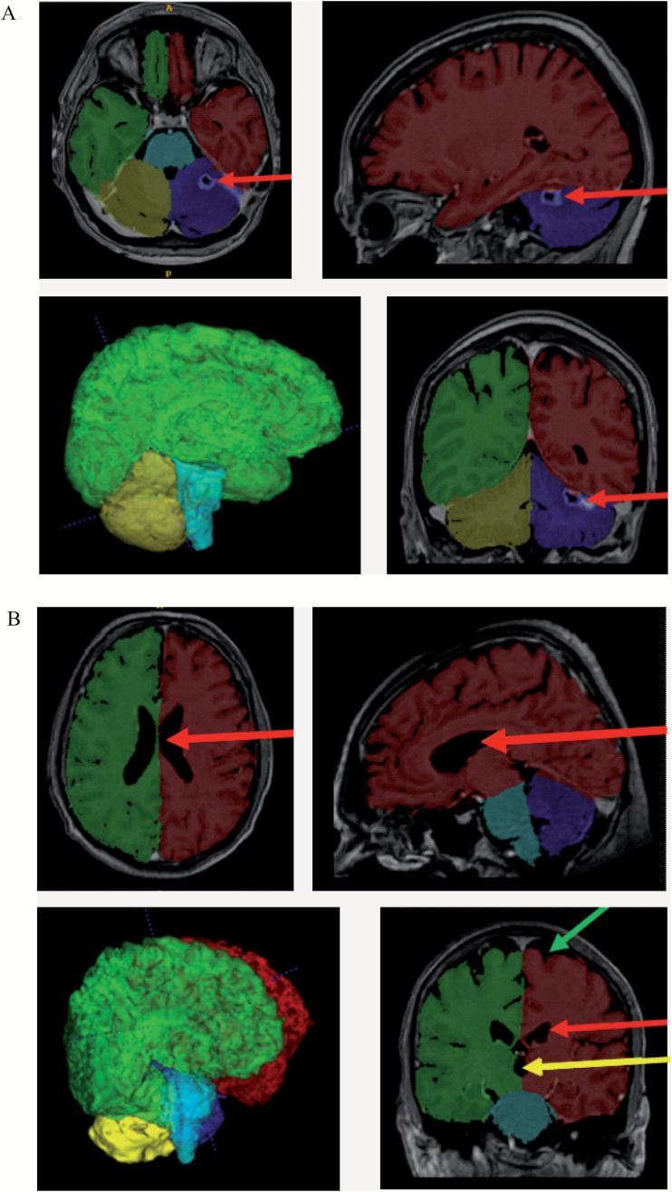
Fig. 1.
Visualization of the results of the volBrain segmentation process with ITK-SNAP. (A) MRI scan of a 45-year-old woman 129 days after commencing WBRT. VolBrain correctly identified the resection cavity in the left cerebellar hemisphere as not being brain tissue (red arrow). The image in the lower-left corner depicts a 3-dimensional reconstruction of the segmentation result created by volBrain. (B) MRI scan of a 68-year-old man 96 days after commencing WBRT. VolBrain exactly differentiated between brain tissue on the one hand and cerebrospinal fluid in the ventricles (third ventricle: yellow arrow; lateral ventricles: red arrow) and subarachnoid space (green arrow) on the other. Description of the segmentation labels: red, left cerebral hemisphere; green, right cerebral hemisphere; blue, left cerebellar hemisphere; yellow, right cerebellar hemisphere; turquoise, brainstem; gray tones, area identified as nonbrain tissue.

