Fig. 1.
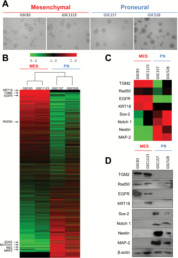
Phenotypic and molecular heterogeneity of glioma stem cells (GSCs). (A) Morphology of mesenchymal (MES) and proneural (PN) GSCs in sphere culture—phase contrast microscopy, under 10x objective. (B) Proteomes of MES and PN cell lines. The main validated subtype-specific proteins are indicated (arrows). (C) Differentially expressed proteins extracted from the proteome of MES and PN GSCs. (D) Western blot validation of differentially expressed marker proteins in MES and PN GSC lines.

