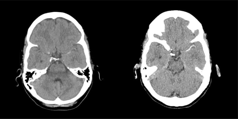
Figure I.

Images of standard NECT (left) and NECT-MIPs (right) of the same patient with an acute ischemic stroke and left M1 occlusion. The NECT-MIPs (right) shows a much more obvious hyper dense vessel sign extending along the left middle cerebral artery when compared to the standard NECT (left).
