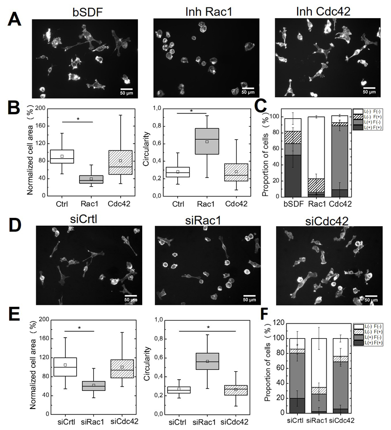
Figure 7.
Effects of drug inhibition or of siRNA-medicated silencing of Rac1 and Cdc42 on bSDF-induced cell spreading and protrusion formation. These effects were assessed 16 h after cell seeding. (A) Fluorescence images (actin staining) of MDA-MB-231 cells seeded onto bSDF films in the presence of Rac1 inhibitor (NSC23766) or Cdc42 inhibitor (ML141). (B) Quantitative analysis of the cell spreading area, circularity and (C) proportion of cells in each depending on their protrusion phenotypes. (D) Representative fluorescence images of cells after silencing using siRNA against Rac1 and Cdc42 and (E) corresponding quantitative analysis (same parameters as in F). At least 50 cells were analyzed for each experimental condition for each experiment. Two independent experiments were performed. *p < 0.05 (Anova one way on ranks).

