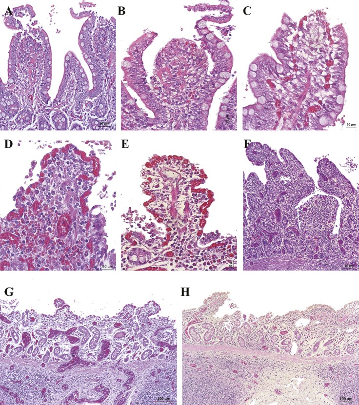
Fig 7. Histopathological images of ileum samples in experimental groups using the Park’s Score (H&E, haematoxyline-eosin staining).
(A) Villi with large subepithelial spaces that have already broken through the epithelium at the tips (Grade 3). There were remnants of epithelial cells in the intestinal lumen. (B and C) Villi with loss of epithelium at the tips and the one that remained on the sides of the villus was detached (Grade 3). Epithelial cells appeared in the intestinal lumen. (D) Villi tips and part of the sides were denuded (Grade 3). Note the congestion and detached epithelial cells. (E) Villi and part of the crypts completely denuded (Grade 4). Note the shortening of the villi, the remains of epithelial cells in the lumen and moderate congestion. (F) Villi and crypts completely denuded. Note the moderate congestion. (G and H) Mucosal intestinal areas without villi (Grade 5). Those villi that still remained on the mucosa were denuded and very short. Note the severe congestion and lymphocytic infiltrate in mucosa and submucosa.

