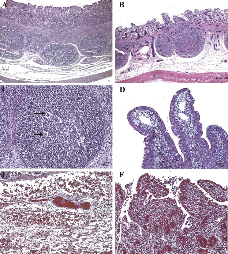
Fig 9. Histopathological images of ileum samples of experimental groups (H&E, haematoxyline-eosin staining).
(A and B) Moderate oedema in submucosa, oedematous Peyer’s patches and severe congestion in mucosa and submucosa was observed in I+ animals. (C) Peyer’s patch with lymphocytic apoptotic forms (arrow). (D) Dilated lymphatic vessels at the villi tips in I- animals. (E) Severe congestion with formation of thrombus and moderate submucosal hemorrhage in I+ animals. (F) Villi without epithelium, with severe congestion and lymphocytic infiltrate in lamina propria observed in I+ animals.

