
Figure 1.
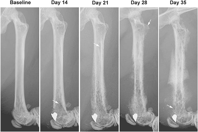
Radiographic images showing disease progression in a mouse model of bone cancer. Bone cancer was induced by drilling a 0.5-mm hole in the center of the trochlear groove of C3H/HeJ male mice (8–9 weeks old) and then injecting and confining 2472 sarcoma cells in the marrow space of the femur. Note that at day 14 post-tumor injection, x-rays show a noticeable tumor-induced bone remodeling which then becomes more severe at days 21, 28 and 35. Day 35 post-tumor injection was the last time point examined in this study.