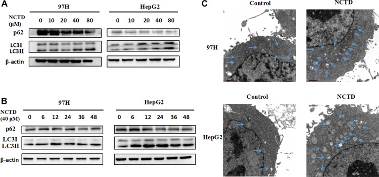
Figure 2. NCTD induces autophagy in HCC cells.
(A) Western blotting analysis of the autophagy-related proteins LC3 and p62 in 97H and HepG2 cells treated with NCTD (0, 10, 20, 40, 80 μM); β-actin was used as a loading control. (B) Western blot of LC3 and p62 in HCC cells exposed to 40 μM NCTD for 0, 6, 12, 24, 36, or 48 h. (C) Quantification of autophagic vacuoles in HCC cells by TEM. Cells were treated with 40 μM NCTD for 24 h.

