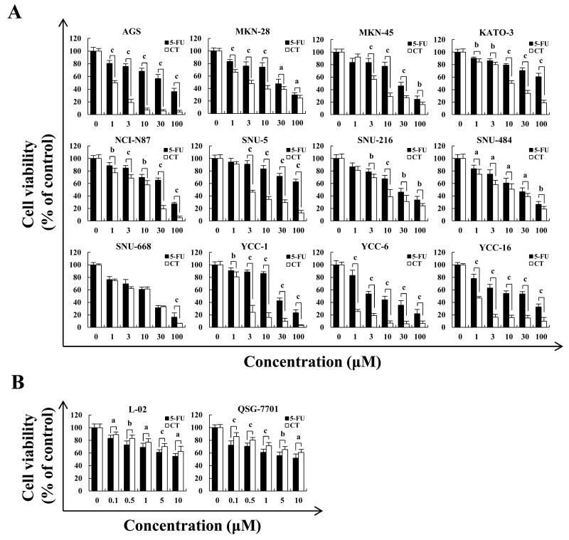
Figure 1. Cytotoxic effects of CT on multiple GC cell lines.
(A) AGS, MKN-28, MKN-45, KATO-3, NCI-N87, SNU-5, SNU-216, SNU-484, SNU-668, YCC-1, YCC-6 and YCC-16 cells were treated with 1, 3, 10, 30 and 100 μM of 5-FU or CT for 24 h, then cell viability was measured by MTT assay. (B) Human liver L-02 and QSG-7701 cells were treated with 0.1, 0.5, 1, 5 and 10 μM of 5-FU or CT for 24 h, then cell viability was measured by MTT assay. Error bars indicate means ± SD of three independent experiments (ap<0.05, bp<0.01, cp<0.001 indicated significant differences).

