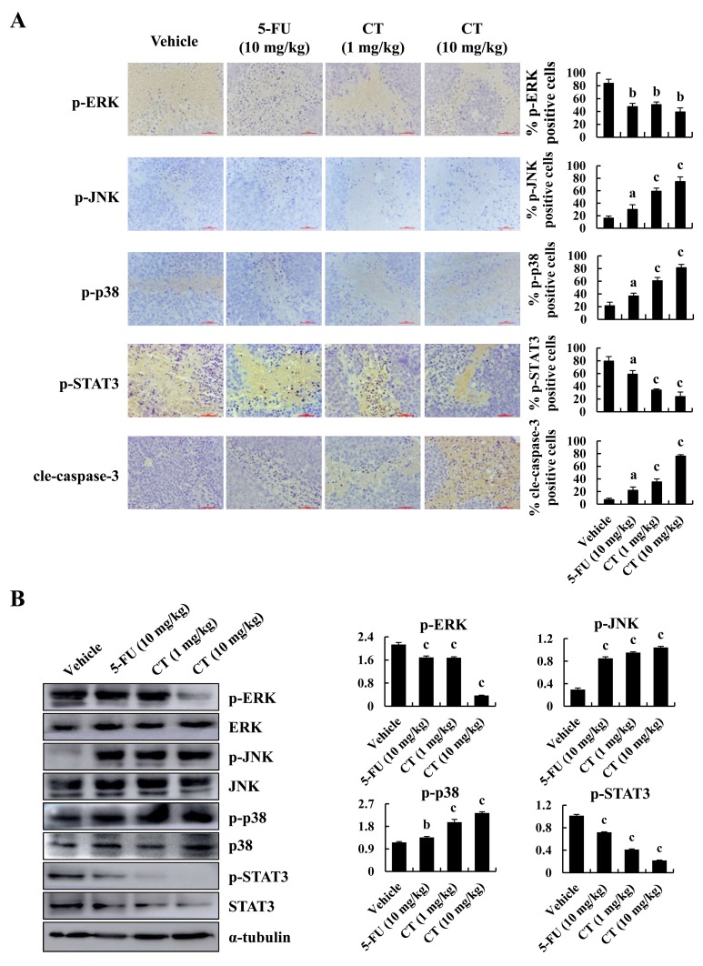
Figure 7. Immunohistochemical detection of key molecules in MAPK signaling pathways in xenograft tumor tissues.
(A) p-ERK p-JNK, p-p38, p-STAT3 and cleaved-caspase-3 expression in tumor tissues was measured by immunohistochemistry under a light microscope. Scale bar 50 μm (ap<0.05, bp<0.01, cp<0.001 indicated significant differences). (B) p-ERK p-JNK, p-p38 and p-STAT3 expression in tumor tissues was measured by western blot (ap<0.05, bp<0.01, cp<0.001 indicated significant differences).

