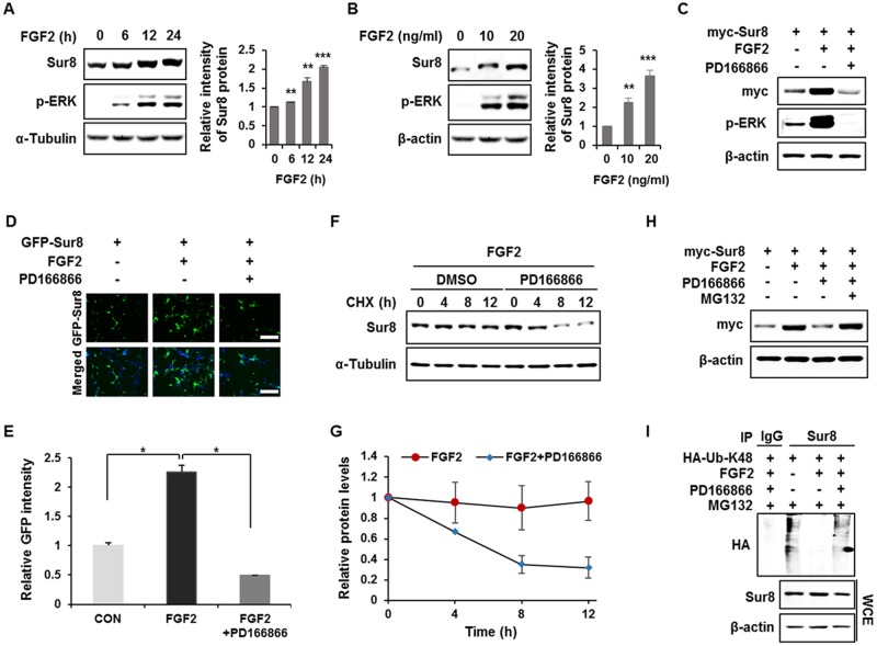
Figure 1. FGF2 stabilizes Sur8 via inhibition of polyubiquitination-dependent proteasomal degradation.
(A, B) HEK293 cells were treated with FGF2 in a time- (A) and dose- (B) dependent manner. (C, D) Cells were transfected with myc-tagged (C) or GFP-tagged (D) Sur8 plasmid. At 24 hours post-transfection, cells were treated with FGF2 (20 ng/mL) for 24 hours, followed by 2-hour pre-treatment with DMSO or a specific FGFR1 inhibitor, PD166866 (100 nM). Representative fields of GFP-positive cells (upper panel) and merged GFP and DAPI (lower panel) are shown (D). Scale bars, 500 μm. (E) Quantified data are shown in graph with mean ± SD from three independent experiments. *P < 0.05. (F, G) Measurement of Sur8 turnover rate by a CHX chase assay. Cells were treated with 50 μg/mL CHX and with FGF2 alone or plus PD166866 for the indicated time periods. The Sur8 protein levels were examined by immunoblot (F) and quantitated (G). (H, I) Cells were transfected with myc-tagged Sur8 (H) or together with HA-tagged K48 ubiquitin (HA-Ub-K48) (I) and treated as indicated for 24 hours followed by 20 μM MG132 treatment for 4 hours before harvesting. Whole cell extracts (WCEs) were immunoprecipitated with an immunoglobulin G (IgG) control or Sur8 antibody (I). WCEs were subjected to immunoblot analysis using the indicated antibodies (A-C, F, H-I).

