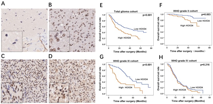
Figure 2. Immunohistochemical staining of HOXD4 in glioma and survival analysis of 453 patients by Kaplan-Meier method.
(A-D) FFPE tissues with HOXD4 expression of normal brain, gliomas of WHO grade II, III and IV respectively (×200, scale bars 50μm), the left bottom of the picture is the enlarged version (×100). (E-H) Overall survival curve by Kaplan-Meier method in cohorts of total glioma patients (p<0.001), glioma WHO grade II (p=0.003), III (p<0.001) and IV (p=0.216) respectively.

