. 2017 Dec 27;6:e33442. doi: 10.7554/eLife.33442

Figure 4. Validation of the CENP-N:CENP-ANCP complex.

(A) In vitro binding assay probing the interaction of GST-CENP-N1-212 immobilized on solid phase with CENP-ANCP. (B) Fluorescence microscopy analysis comparing localization at human kinetochores (U2OS osteosarcoma cells) of a wild-type CENP-N-mCherry fluorescent reporter and of its mutant variants. (C) Quantification of localization of the mCherry constructs in B normalized to CREST. The same concentrations of transiently transfected plasmids were compared. Error bars represent SD.

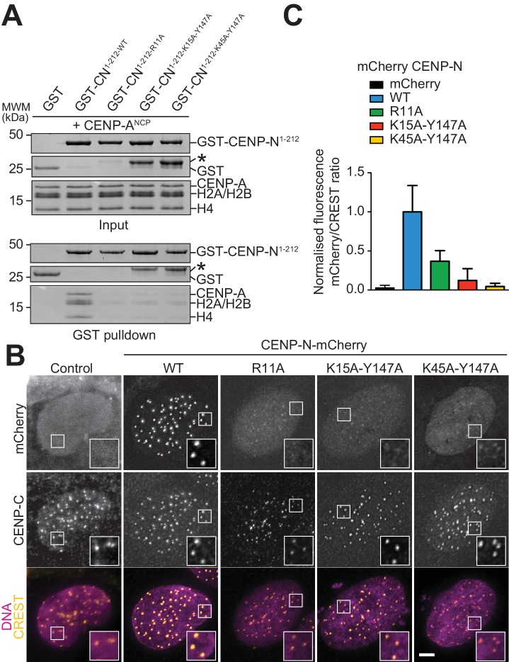
Figure 4.

Figure 4—figure supplement 1. Characterization of CENP-N mutants in solid phase and cell assays.

Figure 4—figure supplement 1.

(A) In vitro binding assay probing the interaction of wild type and mutant GST-CENP-N1-212 immobilized on solid phase with CENP-ANCP. (B) Fluorescence microscopy analysis comparing kinetochore localization of a wild-type CENP-N-mCherry fluorescent reporter and of the indicated mutant variants in U2OS cells. The same concentrations of transiently transfected plasmids were compared.
Figure 4—figure supplement 2. Characterization of CENP-N mutants in competition gel shift assays.

Figure 4—figure supplement 2.

(A–B) Gel shift assays with differentially labelled CENP-ANCPs (red) and H3NCPs (green) at 125 nM in presence of the indicated CENP-N wild type and mutant species (1 µM). The two individual channels are shown below. The nature of all slow-migrating species formed upon binding of CENP-N and its mutants to nucleosomes is unknown, but we suspect they represent higher order oligomerization. Note that mutants affecting the CENP-A L1 loop-binding interface of CENP-N affect CENP-ANCPs binding more than H3NCPs binding, likely because the latter is solely based on the intact DNA-binding interface of CENP-N (see text for additional details).