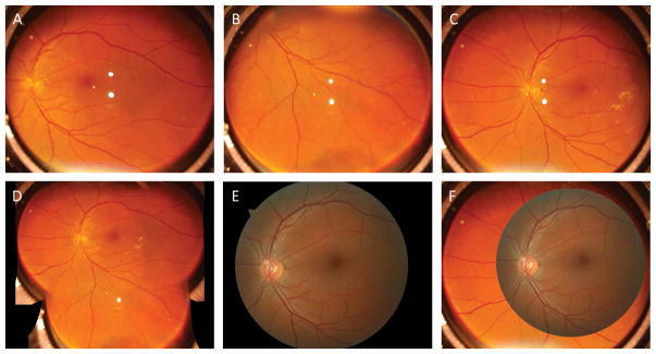
Figure 3.
(A–C) Representative single-shot images captured from a 41 years old age subject. The subject has no reported eye diseases. (D) Montage of single-shot images in Figs. 3A–C. (E) Representative fundus image from the same subject collected with a clinical fundus camera (Zeiss, Cirrus Photo 800), which has a single-shot FOV of 45° external angle, corresponding to 67.5°. (F) Overlap of images of Fig. 3C and Fig. 3E for FOV comparison.

