Figure 2.

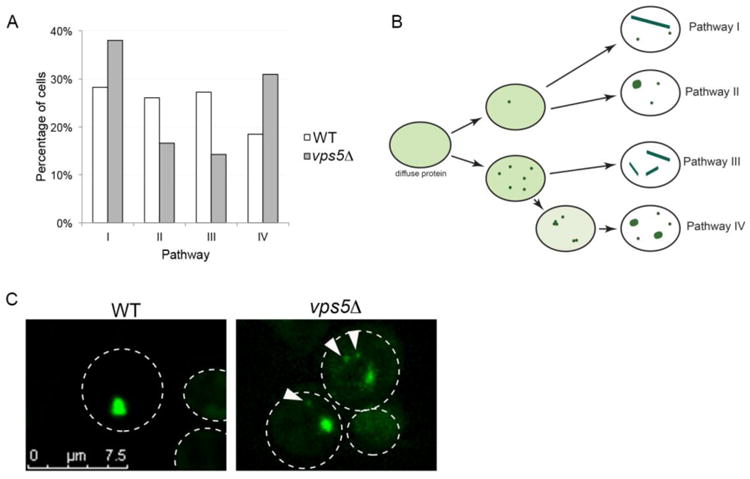
Sup35PrD-GFP forms additional small anomalous aggregates during prion induction. A. Sup35PrD-GFP was overexpressed in vps5Δ cells for 18 hours, and then imaged using 8-well glass slides for an additional 6-12 hours by 4D microscopy. Because of the reduced aggregate formation in vps5Δ, we selectively captured fields of cells in which in the initial stages of early foci formation could be captured. Of the 149 cells imaged, we were able to view aggregate formation in 33 cells (17 cells in G1, and 16 cells in G2/M phase). We followed the formation of early foci into larger aggregates and categorized them into four distinct pathways previously characterized for wildtype cells by Sharma et al., 2017. Statistical analysis using Chi-square goodness of fit tests indicate that while wildtype cells have an equal probability for each pathway, vps5Δ cells do not (p < 0.05). B. Diagrammatic representation of the four pathways in vps5Δ strains. While the pathways were similar between wildtype and vps5Δ strains, we noticed small anomalous aggregates, many of which were mobile, associated with pathways I, II, and IV in vpsΔ. C. Representative images of wildtype (WT) and vps5Δ strains are shown. Arrows indicate small anomalous aggregates. All images were subjected to 3D deconvolution using Autoquant deconvolution algorithms (Media Cybernetics) and are shown as maximum projection images.
