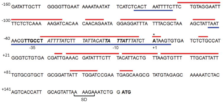
Fig. 4. LeuO and H-NS bind to overlapping DNA sequences at the vieSAB promoter.
The vieSAB TSS (base +1) was determined by 5′-RACE and is indicated with an asterisk. Sequences matching −10 and −35 promoter elements are shown in bold font. The vieS start codon is shown in bold font preceded by the putative Shine-Dalgarno (SD) ribosome binding site. Red lines above the sequence represent regions protected by H-NS on both strands determined by DNase I footprint analysis. Blue lines below the sequence indicate regions protected by LeuO on both strands. A sequence matching the E. coli LeuO binding motif (Dillon et al., 2012) located within the LeuO protected region is indicated in italics.

