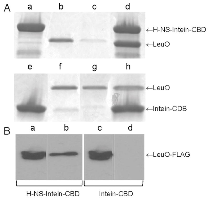
Fig. 6. Interaction between H-NS and LeuO.

A. Fifty μL of chitin beads coupled to an H-NS-intein-CBD fusion (Wang et al., 2012a) (lane a) or to the intein-CBD moiety alone (lane e) were reacted with 20 μg of purified LeuO (lanes b,f). Pull-down was conducted using the binding buffer used for EMSA and Costar Spin-X centrifuge tube filters. Lanes c and g, flow through fractions; lanes d and h, pulled-down fractions. Proteins were separated by SDS-PAGE and stained with Coomassie brilliant blue. B. Fifty μL of chitin beads coupled to an H-NS-intein-CBD fusion (lanes a and b) or to the intein-CBD moiety alone (lanes c and d) were reacted with 0.4 mg of total proteins from a lysate of strain O395ΔlacZΔleuO containing plasmid pUC-rrnB-leuO-FLAG. Lanes a and c, cell lysate input; lanes b and d, pulled-down fractions. LeuO-FLAG was detected by western blot using the anti-FLAG M2-peroxidase monoclonal antibody.
