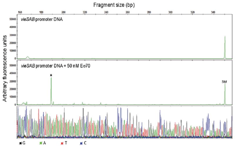
Fig. 7. In vitro transcription of vieSAB DNA.
A vieSAB DNA template spanning nucleotides −160 to +392 relative to the TSS was incubated 1 h at 37○C with or without Eσ70 (negative control). The resulting RNA products were purified and annealed with primer HEX-VieS1 complementary to the vieS open reading frame and extended as described in the methods section. The vieSAB transcription start site is indicated with an asterisk (*). A HEX-labeled DNA standard (Std) was added to each reaction before capillary electrophoresis analysis. The signal from each electropherogram peak is reported as arbitrary fluorescence units along the y axis and the transcript length in base pairs (bp) is reported at the top of the panel. The resulting nucleotide sequence corresponds to the template strand.

