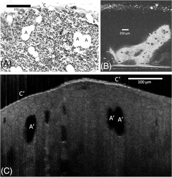
FIGURE 2.

Comparison between (A) hematoxylin-eosin slides (original magnification ×100), (B) en face optical frequency domain imaging (OFDI), and (C) cross-sectional μ-optical coherence tomography (μOCT) images of the normal parathyroid gland. The gland is covered by a delicate capsule (C); its parenchyma is composed by chief cells, oncocyte cells, and adipose cells (A). Adipocytes are easily recognized in OFDI (arrows) and μOCT as a large dark ovoid or round shaped structures (A′). A thin but bright capsule covers the glandular parenchyma (C′).
