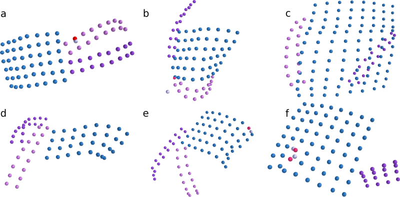
Fig. 4.
Results of the sorting and labeling algorithm are shown for six of twelve subjects with subdural grids implanted. The algorithm is robust to identifying correct electrode configurations even when grids are substantially folded (e.g., across the cortex). a–f Correctly sorted electrodes are displayed by grid color in blue, purple, and pink. Incorrectly sorted electrodes are displayed in red. True electrodes that were not identified correctly by the sorting algorithm are displayed in light gray. Additional candidate electrode locations correctly identified as noise are not shown

