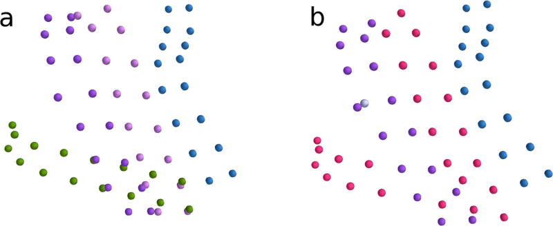
Fig. 6.
a True grid configuration of one subject with overlapping grids. Each color represents a distinct grid. b Results of sorting and labeling algorithm for the same subject. Correctly sorted electrodes are shown in blue and purple. True electrodes that were not identified correctly by the sorting algorithm are displayed in light gray. The improperly omitted electrodes, belonging to two grids which were not identified, are shown in red

