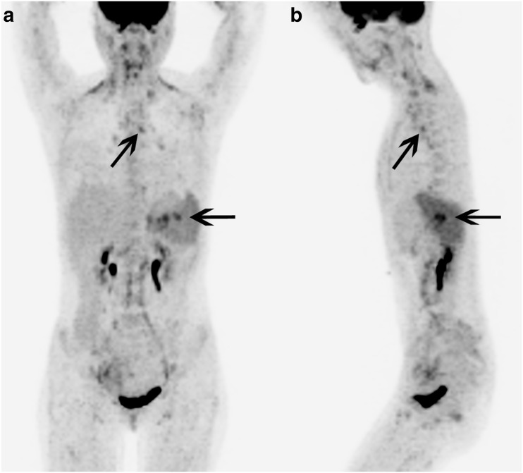
Fig. 1.
Patient 1. 18F-FDG PET/CT maximum intensity projection (a) anterior and (b) left lateral images show diffuse mildly 18F-FDG avid lymphadenopathy with several foci of 18F-FDG uptake in the AP window (diagonal arrows), and the right lower paratracheal and subcarinal regions, as well as several foci of intense 18F-FDG uptake in the region of the splenic hilum (left-pointing arrows). Histological evaluation of these areas confirmed Kikuchi-Fujimoto disease. There is also diffusely increased 18F-FDG uptake in the entire spleen

