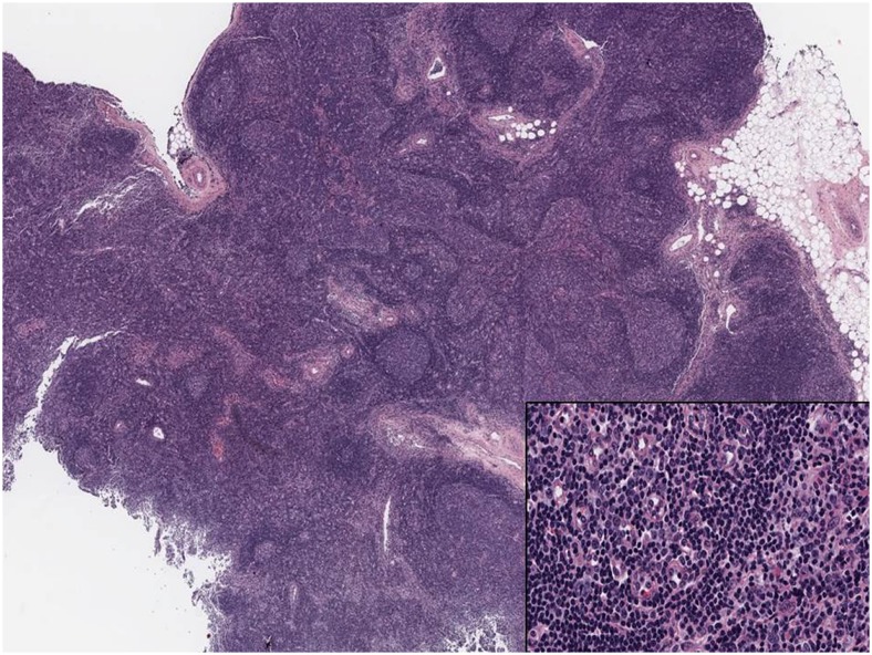
Fig. 6.
Patient 2. Immunohistochemical evaluation of a left axillary lymph node showing follicular hyperplasia with enlarged germinal centers, intact mantle zones and follicles of variable size and shape. Inset: High-power view of the germinal center with multiple tingible body macrophages and apoptotic bodies. There was no evidence of lymphoma

