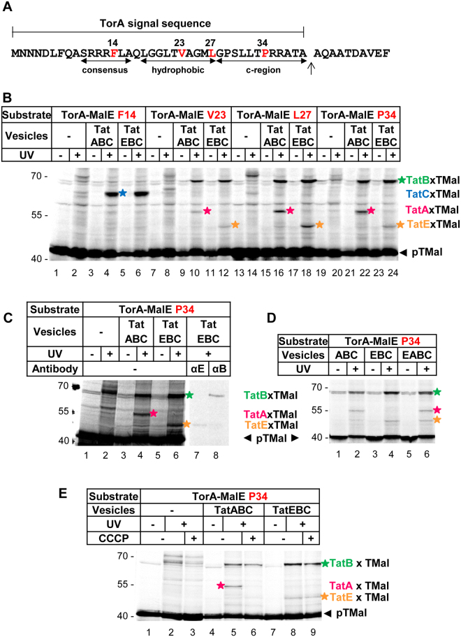
Figure 1.
TatE interacts with the signal sequence of a Tat substrate. (A) Primary structure of the TorA-signal sequence. The twin-arginine consensus motif, the hydrophobic core and the c-region are indicated. Amino acid residues substituted by the photo-crosslinker p-benzoyl-L-phenylalanine (Bpa) are labeled in red. (B–E) The model Tat substrate TorA-MalE335 (TMal) was in vitro synthesized and radioactively labeled. Bpa was incorporated into the signal sequence at the indicated positions (red). After incubation with inverted inner membrane vesicles containing the indicated Tat components, crosslinking was induced by UV-irradiation (UV). The samples were resolved by 10% SDS-PAGE and analyzed by phosphorimaging. Adducts between the substrate and TatC (blue star), TatB (green star), TatA (magenta stars) and TatE (orange stars) are indicated. (C) Adducts between TMal, TatE and TatB were confirmed by immuno-precipitation using antisera against TatE and TatB. The eight lanes are derived from a single gel after excising a piece between lanes 6 and 7. (D) In vesicles containing TatE alongside TatA, the signal peptide cross-links to both Tat proteins. (E) Dissipation of the proton-motive force by 0.1 mM CCCP abolishes contacts with TatA but not with TatE.

