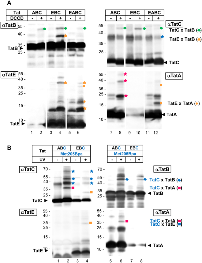
Figure 6.
TatE-specific interactions with TatA, TatB and TatC. Western blot analysis of crosslinking experiments with inverted inner membrane vesicles containing the indicated Tat subunits (TatABC, EBC, EABC). After crosslinking, the samples were resolved using 9% Tricine-SDS-PAGE and detected by immunoblotting using antibodies against TatB (αTatB), TatC (αTatC), TatE (αTatE) and TatA (αTatA). (A) Crosslinking using N,N′-dicyclohexylcarbodiimide (DCCD). After incubation with DCCD higher molecular bands representing TatB-TatC adducts (green diamond), TatE-TatB adducts (orange triangle), and TatE-TatA adducts (orange dots) were detected. Indicated are the positions corresponding in size to TatC dimers (blue star), TatE dimers (orange star) and TatA oligomers (magenta stars). Lanes are derived from four individual gels. The gaps mark the excision of single unrelated lanes. (B) Crosslinking using p-benzoyl-L-phenylalanine (Bpa). During vesicle preparation the crosslinker was incorporated into TatC at position 205 (Met205Bpa). Bpa-crosslinking was induced by exposing the vesicles to UV irradiation. UV-dependent higher molecular bands were identified via their immuno-reactivities as TatC oligomers (blue star), TatB-TatC adducts (blue diamond), TatC-TatA adducts (magenta square) and TatC-TatE adducts (orange square).

