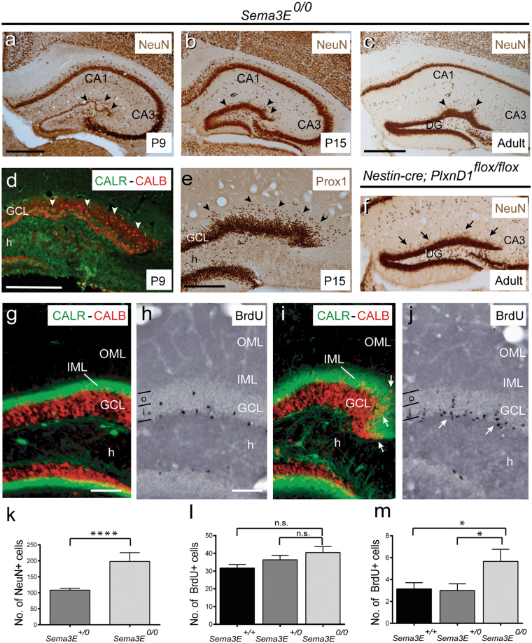
Figure 4.
Examples of α-NeuN immunostaining in the hippocampus proper and dentate gyrus of Sema3E0/0 (a–c) and Nestin-cre; PlxnD1flox/flox (f) mice at different postnatal stages. Note the presence of numerous NeuN-positive cells in the molecular layer in absence of Sema3E/PlexinD1 signalling (arrowheads), especially in Sema3E-deficient mice. Also note the presence of several waves of the suprapyramidal blade of dentate gyrus. (d) Double immunolabeling of Calretinin (green) and Calbindin (red) in the dentate gyrus of Sema3E0/0 mice at P9. Note the presence of numerous ectopic Calbindin-positive neurons in the IML of Sema3E0/0 mice (arrowheads). (e) Prox-1 immunolabeling in the dentate gyrus of Sema3E0/0 mice at P15 shows the presence of numerous Prox-1-positive granule cells in the molecular layer (arrowheads). (g,i) Double immunolabeling of Calretinin (green) and Calbindin (red) in the dentate gyrus of control (g) and Sema3E0/0 adult mice (i). Note the presence of numerous ectopic Calbindin-positive neurons in the IML of Sema3E0/0 mice (arrows in i). (h–j) Examples of BrdU-labeled neurons in the granule cell layer of control (h) and Sema3E0/0 (j) mice. Numerous BrdU-positive cells forming columns in the granule cell layer can be seen in mutant mice (arrows in j). (k) Histogram illustrating the number of NeuN-positive cells in the IML of the suprapyramidal blade of the dentate gyrus in Sema3E+/0 and Sema3E0/0 mice. (l–m) Histograms illustrating the number of BrdU-positive cells counted in the whole granule cell layer (l) and in the outer portion of the granule cell layer of the dentate gyrus (labeled as ‘o’) (m) in Sema3E+/+, Sema3E+/0 and Sema3E0/0 mice. Asterisks indicate statistical differences between groups. *P ≤ 0.05; **P ≤ 0.01; ***P ≤ 0.001; ****P ≤ 0.0001. ANOVA Bonferroni post hoc test. Abbreviations as in Figs 1–3 and i = inner portion of the granule cell layer; IML = inner molecular layer; o = outer portion of the granule cell layer and OML = outer molecular layer. Scale bars: a = b = 500μm; c = f = 500 μm; d = 200 μm; e = 200 μm; g and h = 150 μm pertains to i and j respectively.

