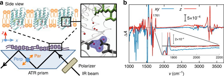
Fig. 2.
Experimental polarization-resolved spectra. a In the experimental set-up, the purple membranes are oriented in the xy plane and the proton pumping direction is along z. The black arrow indicates the direction of the probing IR light, which is polarized parallel or perpendicular with respect to the plane of incidence. The inset shows a zoom-in of the crystal structure of bacteriorhodopsin70, with three water molecules highlighted in blue, which presumably cause the continuum band22, 25. b Experimental IR light-minus-dark difference spectra calculated along the xy and z directions over the whole measured range between 1000 and 3800 cm−1. In the inset, the enlarged difference absorption spectra in the relevant wavenumber range 1700–2200 cm−1 are shown together with the (positive) band at 1761 cm−1 of the C=O stretching vibration of D85

