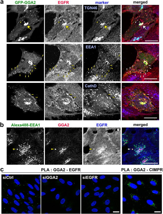
Figure 4.
GGA2 colocalizes and interacts with EGFR in vivo. (a) ARPE-19 cells overexpressing GFP-tagged GGA2 were fixed and double immunofluorescence staining was performed using antibodies against EGFR (red) and TGN46, EEA1, or cathepsin D (blue). Arrows and arrowheads indicate colocalization of three signals in the TGN and peripheral structures, respectively. Bar, 20 μm. (b) ARPE-19 cells were fixed for triple immunofluorescence analyses using antibodies against Alexa488-EEA1 (green), endogenous GGA2 (red) and EGFR (blue). Arrowheads indicate colocalization of three signals. Bar, 20 μm. (c) ARPE-19 cells transfected with siCtrl, siGGA2, or siEGFR were processed for PLA to detect GGA2-EGFR or GGA2-CIMPR interaction (green). Nuclei were stained with DAPI (blue). Bars, 20 μm.

