Figure 7.
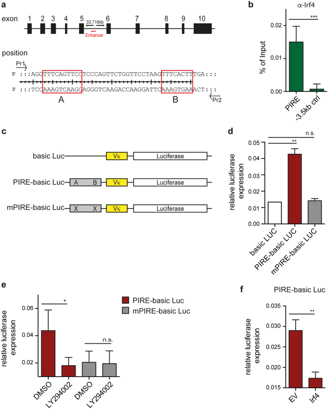
Irf4 directly represses Pax5 expression. (a) Schematic overview showing the location of the Pax5 enhancer region within the Pax5 gene locus. (b) Irf-4 ChIP from cells of a SLP-65-deficient pre-B cell line. Amplified by qPCR was the enhancer region of Pax5 within exon 5 containing Irf4-binding sites (A and B). As control, amplification of a region 3.5 kb upstream of Pax5 enhancer was chosen. Both samples were normalized to mock control. (c) Schematic overview of the luciferase expression vector harboring a Vκ promoter and the respective Pax5 enhancer region containing or lacking the Irf4 binding sites. (d–f) WEHI cells were electroporated to introduce the empty vector (Vκ + Luciferase) or the indicated constructs containing or lacking the Irf4 binding sites. Expression of luciferase was equalized to the rLUC (Renilla) expression in each sample of co-transfected WEHI cells. In (e), WEHI cells were treated for 16 h with LY294002 upon electroporation and in (f), WEHI cells were co-transfected with either EV or with a vector encoding Irf4. Data are representative of at least 3 independent experiments and luciferase expression was determined using the Dual-Luciferase Reporter Assay System.

