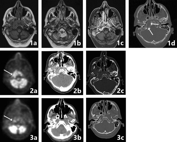
Fig. 6.
SBO crossed extension. A 64-year-old patient presented with clinical signs of a mastoid abscess. After initial successful surgery and antibiotic therapy, the patient presented 4 weeks later with progressive cranial deficit of the X and XI nerve. Imaging at that time was performed with CT and MRI in a referring hospital, with PET-CT imaging at 1 month (image 2a–c) and at 6 months follow-up. MRI with T1-w, T2-w, contrast-enhanced T1w-fs images (image 1a, b, c) and CT (image 1d) are shown. There is marked hypointensity at the bone marrow of the right skull base, the clivus and to a lesser extent of the left skull base, with involvement of the subtemporal soft tissues, the masticator space at right side, the prevertebral tissues and nasopharyngeal wall, with crossed extension to the left side. T1 FS post-gadolineum scan illustrates the extent of bone and soft tissue involvement (image 1c). Additional high-resolution CT shows bone erosion in the corresponding region, especially at the medial part of the skull base, the jugular foramen and the clivus (image 1d arrows). FDG-PET-CT indicates the metabolic activity and spread of the inflammation sites (image 2a arrows) and was used for follow up with a normalization of the FDG avidity after 6 months (image 3a arrows). The healing process is to a lesser extent seen on CT, but there is healing and remodeling of the cortical borders of the clivus (image 3c)

