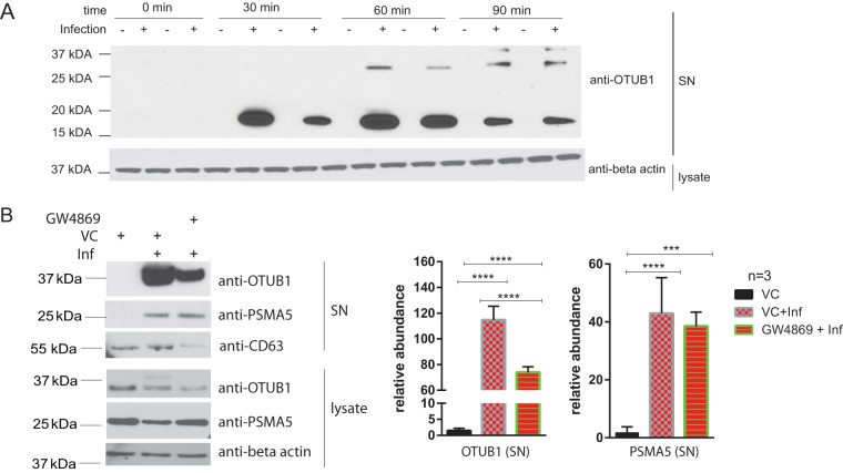
FIG 5.
(A) THP-1 macrophages were infected or left uninfected for 0, 30, 60, and 90 min with S. Typhimurium. Cell culture medium was collected at each time point in duplicates and analyzed by Western blotting with anti-OTUB1 and anti-β-actin antibodies (a loading control for cell lysate). (B) THP-1 macrophages were treated with GW4896 inhibitor (5 μM) or an equivalent volume of DMSO (vehicle control [VS]) for 90 min before infection. Cells were infected (Inf) for 90 min with S. Typhimurium at an MOI of 50:1 or left uninfected. CD63 (exosome marker), OTUB1, and PSMA5 were detected in cell culture supernatant (SN), whereas OTUB1, PSMA5, and β-actin (a loading control) were detected in cell lysate by Western blotting. ImageJ was used to quantify the pixel density of the bands (n = 3). A Student t test was used for statistical analysis. The results are displayed as relative abundances on graphs. P values are indicated as follows: *, P ≤ 0.05; **, P ≤ 0.01; ***, P ≤ 0.001; and ****, P ≤ 0.0001.

