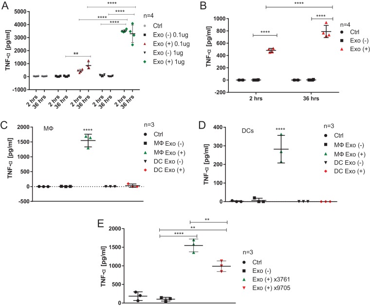
FIG 7.
Exosomes derived from infected macrophages stimulate release of TNF-α from naive macrophages. (A) RAW 264.7 mouse macrophages were infected with wild-type S. Typhimurium (UK-1; MOI of 5:1) or left uninfected. Exosomes were collected from cell culture supernatants at 2 and 36 hpi and their concentration was established by BCA protein assay. Naive RAW 264.7 macrophages were treated with PBS (Ctrl) or with 0.1 or 1 μg of exosomes derived from infected and uninfected macrophages. After 24 h, the cell culture supernatant was collected, and TNF-α release was measured by ELISA. Results from four biological replicates are shown. Two-way ANOVA test was used to establish statistical significance, which was done in conjunction with multiple testing correction (Tukey). Exo(+), exosomes derived from infected macrophages; Exo(−), exosomes derived from uninfected macrophages. (B) Mouse BMDMs were treated with 1 μg of exosomes derived from uninfected or S. Typhimurium-infected RAW 264.7 macrophages (2 and 36 hpi, MOI of 5:1). The cell culture supernatant was collected after 24 h, and TNF-α release was measured by ELISA. Four biological replicates are shown. One-way ANOVA test with Tukey's multiple testing correction was used to establish statistical significance. (C) Exosomes were isolated from uninfected and S. Typhimurium-infected BMDMs or BMDCs (2 hpi, MOI of 5:1). Naive BMDMs were treated with these exosomes or PBS (Ctrl) for 24 h, and TNF-α release was then measured by ELISA. The results of three biological replicates are shown. One-way ANOVA test with Tukey's multiple testing correction was used to establish statistical significance. (D) Exosomes were isolated from uninfected and S. Typhimurium-infected BMDMs or BMDCs as in panel C. Naive BMDCs were treated with these exosomes or PBS (Ctrl) for 24 h, and the TNF-α release was then measured by ELISA. The results of three biological replicates are shown. One-way ANOVA test with Tukey's multiple testing correction was used to establish statistical significance. (E) THP-1 macrophages were infected with wild-type S. Typhimurium (UK-1 χ3761; MOI of 5:1), infected with ΔpagL7 ΔpagP81::Plpp lpxE ΔlpxR9 S. Typhimurium (UK-1 χ9705; MOI of 5:1), or left uninfected. Exosomes were purified from cell culture supernatant at 2 hpi. Naive THP-1 macrophages were treated with PBS (Ctrl) or with 1 μg of exosomes derived from infected and uninfected macrophages. After 24 h, the cell culture supernatant was collected, and TNF-α release was measured by ELISA. The results of three biological replicates are shown. One-way ANOVA test with Tukey's multiple testing correction was used to establish statistical significance. P values are indicated as follows: *, P ≤ 0.05; **, P ≤ 0.01; ***, P ≤ 0.001; and ****, P ≤ 0.0001.

