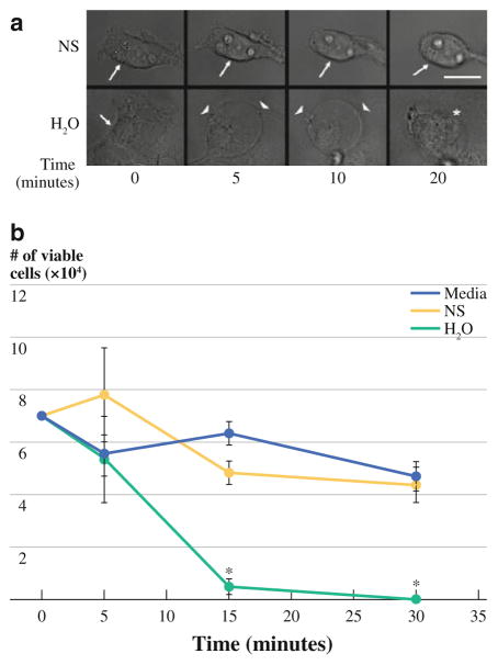
FIG. 1.
a Representative photomicrographs obtained from video capture demonstrate the loss of adherence of CT26 cells exposed to saline treatment (NS) (no cell membrane, cytosol, or nuclear changes noted). CT26 cells exposed to H2O show immediate morphological changes with rounding, cell membrane bleb formation, loss of cytosol, and eventual loss of cell and nuclear membrane integrity within 10–20 min (scale bar = 30 μM; arrow = cell membrane, arrowhead = membrane rounding with bleb formation, asterisk = membrane disruption). b CT26 cell viability was minimal after 15 min of exposure to H2O and differed significantly from NS- or media-treated cultures (P <0.05)

