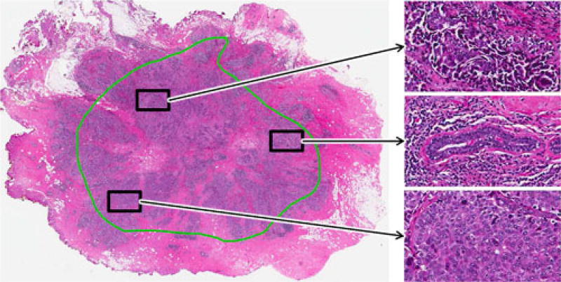
Fig. 2.
FOVs taken from a single histopathology slide illustrate the high level of intratumoral heterogeneity in ER+ BCa. The green annotation represents invasive cancer as determined by an expert pathologist. Note the disorganized tissue structure of FOVs with higher malignancy (top, bottom) compared to the FOV containing benign tissue (middle).

