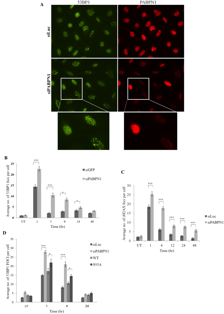
Figure 5.
PABPN1 presence and phosphorylation are required for timely dissolution of NCS-induced 53BP1 nuclear foci. (A) U2-OS cells transfected with siLuc or siPABPN1 were treated with 3.5 ng/ml NCS, and co-immunostaining of PABPN1 and 53BP1 was carried out 8 h later. Note the striking difference between PABPN1-positive and -negative cells with regard to presence of 53BP1 foci. (C) Average counts of 53BP1 foci at various time points after treatment with 3.5 ng/ml NCS in PABPN1-proficient and -deficient cells (average of 100 cells). Bars represent SEM. Only cells negatively stained for PABPN1 were considered PABPN1-deficient. (C) Average counts of γH2AX nuclear foci at various time points after irradiation with 1 Gy IR in PABPN1-proficient and –deficient cells (average of 100 cells). Bars represent SEM. Only cells negatively stained for PABPN1 were considered PABPN1-deficient. (D) Similar analysis as in (B) of cells in which endogenous PABPN1 was depleted and cells in which it was replaced by ectopic, WT or S95A mutant PABPN1. The data represent five sets of experiments for 53BP1 and two sets for γH2AX. Error bars represent SEM. Statistical analysis was based on Student's t test.

