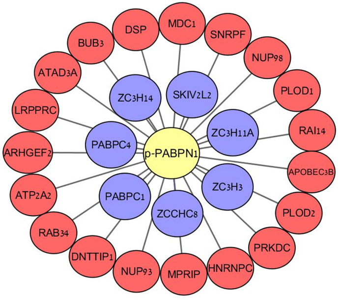
Figure 8.
Protein-protein interactions of phospho-PABPN1 following induction of DNA damage. U2-OS cells were treated with 50 ng/ml NCS for 30 min and cell lysates were used for immunoprecipitation using antibodies against pPABPN1 or total PABPN1. The immune complexes were subjected to mass spectrometry analysis. The specific interactors of pPABPN1 are presented. Red nodes – proteins that precipitated only with phospho-PABPN1. Blue nodes—proteins that precipitated with both phospho- and total-PABPN1.

