
Figure 1.
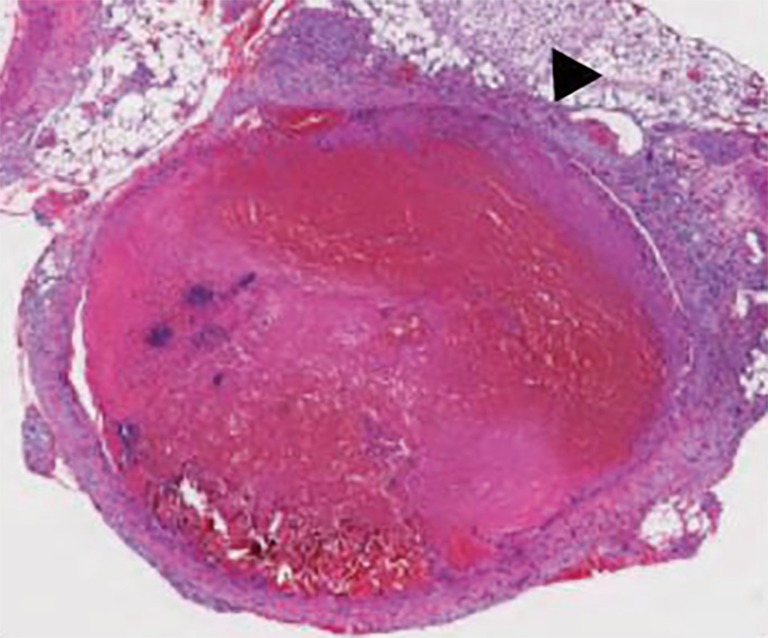
Image is 100× in magnification. Hematoxylin and eosin staining of a 5 micron section of a femoral vein fixed in 10% formalin and embedded in paraffin. The image demonstrates marked dilation of the vein containing a thrombus that is 3 days old. Black arrowhead indicates the vein wall.