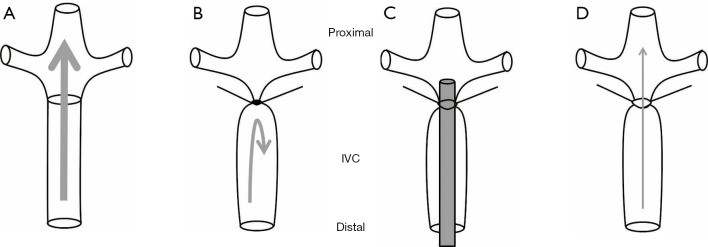
Figure 1.
Diagram of venous stasis and stenosis models in the IVC. (A) Normal IVC blood flow; (B) applying a ligature distal to occlude the renal veins creates stasis; (C) placing a spacer on top of the IVC followed by looping a ligature around the IVC and the spacer; (D) subsequently removing the spacer will create venous stenosis with partial IVC occlusion. IVC, inferior vena cava.

