
Fig. 4.
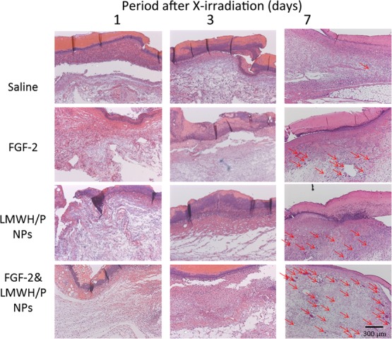
Each photomicrograph of the saline (control)-, FGF-2-, LMWH/P NPs- and FGF-2&LMWH/P NPs-administered wounds in the X-irradiated groups on Days 1, 3 and 7 is representative of six preparations. The red arrows indicate blood vessels with several erythrocytes.