
Fig. 5.
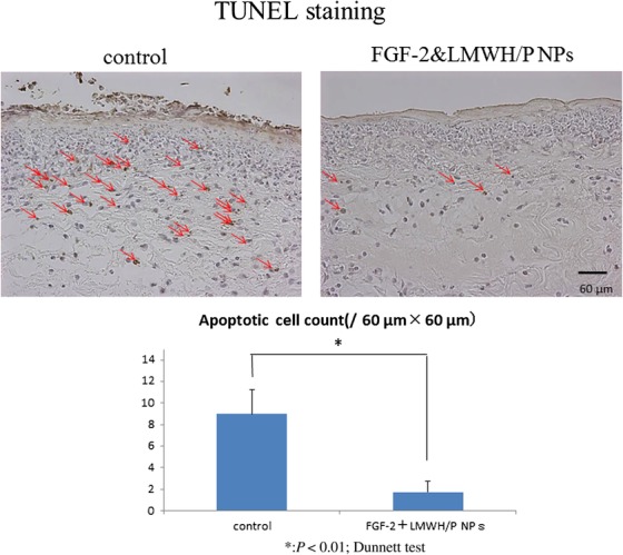
TUNEL-staining of X-irradiated skin. Control- and FGF-2&LMWH/P NPs-administered X-irradiated wounds were TUNEL-stained 24 h after wound creation. The red arrows indicate TUNEL-stained spots. Each photomicrograph is representative of eight preparations. The number of TUNEL-stained spots is significantly higher in saline (control)-administered X-irradiated wounds than in FGF-2&LMWH/P NPs-administered X-irradiated wounds. (P < 0.01, n = 8).