
Figure 5.
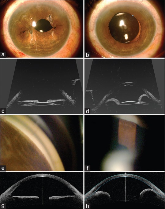
Comparative images of both eyes of a case of angle-closure glaucoma with plateau iris syndrome and cataract extraction (left column = left eye with single-pass four-throw; right column = right eye with no single-pass four-throw). (a) Postoperative image of the left eye with single-pass four-throw and cataract extraction. (b) Postoperative image of the right eye with only cataract extraction done. (c) Ultrasound biomicroscopy denotes open angles with flat iris tissue. (d) Ultrasound biomicroscopy denotes iris bombe with peripheral anterior synechia. (e) Gonioscopy shows open angles. (f) Gonioscopy shows closed angles. (g) Anterior segment optical coherence tomography shows open angles. (h) Anterior segment optical coherence tomography shows angle closure with iris bombe