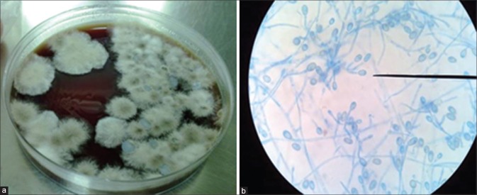
Figure 1.
(a) Chocolate agar showing dirty white cottony colonies on inoculation of corneal scraping material. Growth was seen after 3 days of incubation at 25°C. (b) Lactophenol cotton blue staining showing septate hyphae, ovoid conidia (as shown by the microscope pointer) with truncated bases suggestive of Scedosporium apiospermum as visualized using a binocular compound microscope with high power and ×45

