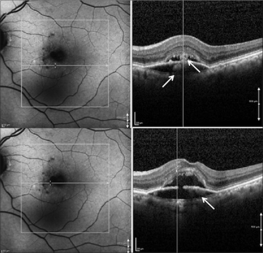
Figure 6.

Same visit as in Figure 5 showing a new hyperautofluorescent halo appearing around the hypoautofluorescent lesion, with a corresponding hyperreflective material both above and below the retinal pigment epithelium (top right and bottom right) on spectral domain-optical coherence tomography scans at different regions
