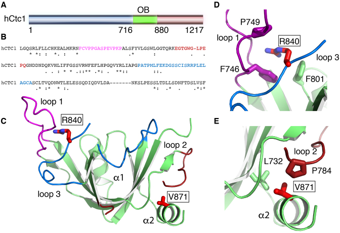
Figure 1.
X-ray crystal structure of a central OB-fold of human Ctc1. (A) Primary sequence of hCtc1. (B) Sequence conservation of hCtc1(OB); the following genes were used for the alignment; Human, zebrafish, mallard, Vitis vinifera, mouse, rat, Spermophilus tridecemlineatus, rabit, killifish, bovine, African bush elephant, orangutan. Color font represents the surface loops of hCtc1(OB). (C) Crystal structure of hCtc1(OB) with surface loops highlighted in color. The disease-associated mutations R840W and V871M are shown in red stick. (D) Zoom-in representation of R840 and the residues interacting with it. (E) Zoom in representation of V871 and the residues interacting with it.

