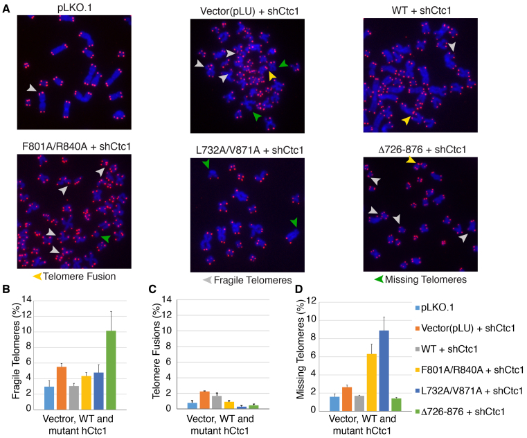
Figure 7.
Metaphase telomere FISH reveals that mutation or deletion of hCtc1(OB) leads to telomeres defects. (A) Telomeric DNA FISH analysis on metaphase spreads in 293T cells stably expressing full length, WT and mutant (double and deletion) hCtc1 proteins. Missing telomere signal, fragile telomeres and telomere fusions are indicated by green, gray and yellow arrowheads respectively. (B) The bar graphs in panels B-D show the various levels of dysfunctional telomeres for the hCtc1 double and deletion mutants. A total of 1000 chromosomes were used for each hCtc1 mutant. Standard deviation for 1000 chromosome measurements for WT and mutant hCtc1 are shown as error bars.

