Fig. 4.

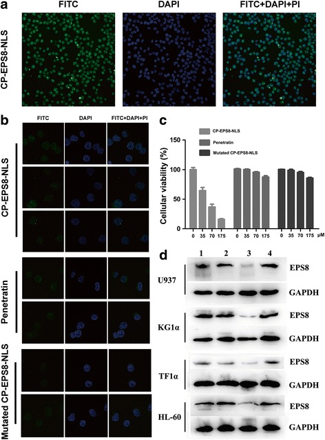
Intracellular distribution of CP-EPS8-NLS in U937 cells. a U937 cells (2 × 10⁵ cells per plate) were transduced with FITC-conjugated CP-EPS8-NLS (40 μM) in 1 mL of culture medium for 2 h and stained with PI and DAPI. Significant green fluorescence could be observed both in the nucleus and cytoplasm. b U937 cells were treated with either mutated CP-EPS8-NLS or penetratin. Green fluorescence was only observed in the cytoplasm. c U937 cells were treated with increasing concentrations of CP-EPS8-NLS, mutated CP-EPS8-NLS, and penetratin. After 24 h, a CCK-8 assay was performed. d Four AML cell lines were treated with 70 μM CP-EPS8-NLS, mutated CP-EPS8-NLS, and penetratin for 12 h and analyzed by western blot. CP-EPS8-NLS significantly decreased the expression of EPS8, while mutated CP-EPS8-NLS and penetratin did not (1. Control; 2. Mutated CP-EPS8-NLS; 3. CP-EPS8-NLS; 4. Penetratin)
