
Figure 2.
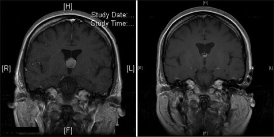
Coronal T1 MR images with gadolinium of patient #2. Left: Discovery MRI showing the 12-mm colloid cyst lesion at the anterosuperior aspect of the third ventricle. Right: 4-year post-aspiration and radiosurgery MR images demonstrating only the colloid cyst capsule remnants