
Fig. 1.
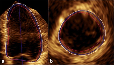
(a) LV apical 4-chamber view and (b) basal short-axis view in ED showed that the Heartmodel software detected the inner and outer extents of the myocardial tissue, i.e., the blood-tissue interface (red line) and the interface of compacted myocardium (white line), which were assigned to slides of 0% and 100%, respectively. The LV endocardial border (blue line) between them was subjectively defined by the operator, and in this case, it was at the slide of 68%