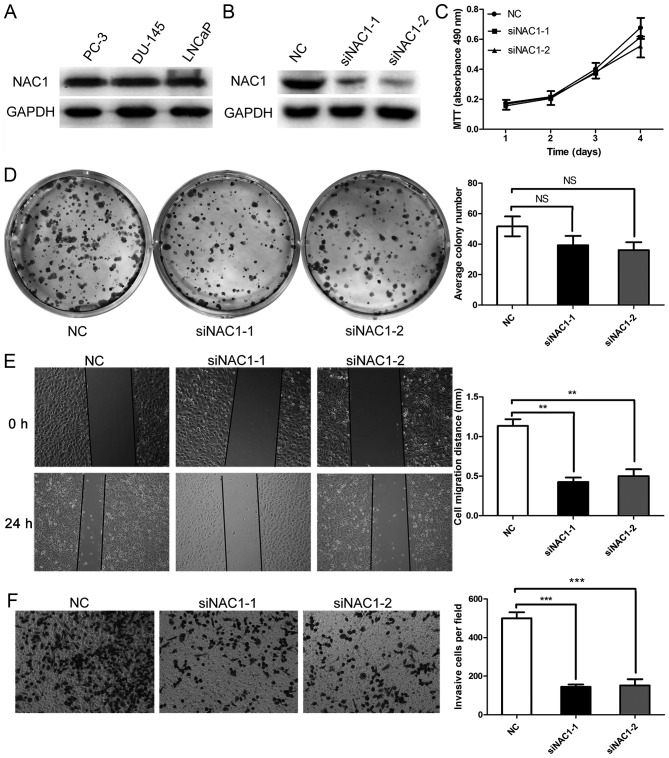
Figure 2.
Silencing NAC1 expression decreased the migratory ability of prostate cancer cells. (A) Western blots showing expression of NAC1 in the highly aggressive prostate cancer cell lines PC-3, DU-145 and LNCaP. (B) Western blots showing a significant reduction of NAC1 protein in DU-145 cells transfected with NAC1 siRNAs (siNAC1-1 and siNAC1-2) compared with those transfected with negative control siRNA (NC). (C) MTT assay showing that siNAC1-1 and siNAC1-2 had no obvious effect on the cell number of DU-145 cells (P>0.05). (D) Detection of cell proliferation by plate colony formation assay in DU-145 cells transfected with NAC1 or NC siRNAs. Representative photographs show the DU-145 cell colonies in 6-well plates on the left. The cell colonies were scored visually and counted using a light microscope, as shown in the graph on the right; the siNAC1-1 and siNAC1-2 transfection groups exhibited no distinct difference in the number of colonies compared with the NC siRNA group (NS P>0.05). (E) Wound healing assay showed that NAC1 silencing affected the migration of prostate cancer DU-145 cells, as shown in the photographs on the left. The comparison of cell migration distance between siNAC1-1 or siNAC1-2 and NC siRNA transfection is shown on the right (**P<0.01). (F) Transwell migration analysis showed that NAC1 silencing affected the migration of prostate cancer cells, as shown in the photograph on the left (magnification, 200x). The quantitative analysis of the migratory ability of prostate cancer cells transfected with NAC1 or NC siRNAs is shown on the right (***P<0.001).

