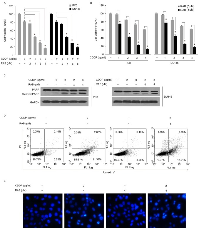
Figure 2.
RAB enhances the cytotoxicity of CDDP. RAB sensitized PC3 and DU145 cells to CDDP-mediated anti-proliferation and apoptosis; (A) PC3 and DU145 cells were treated with different concentrations of RAB (2–8 µM) and a fixed concentration of CDDP (2 µg/ml) for 48 h at 37°C; (B) PC3 and DU145 cells were treated with different concentrations of CDDP (1–4 µg/ml) and a fixed concentration of RAB (4 µM) for 48 h at 37°C as measured by MTT assay. *P<0.05 vs. single treatment with the corresponding concentrations of CDDP. RAB sensitized PC3 and Du145 cells to CDDP-induced apoptosis as measured by (C) PARP cleavage and (D) flow cytometry. (E) Apoptosis in PC3 cells as visualized using DAPI staining. Cells were exposed for 48 h at 37°C to the indicated treatments prior to staining with DAPI for 30 min. CDDP, cisplatin; RAB, Retigeric acid B; PI, propidium iodide; PARP, poly adenosine 5′-adenosine diphosphate ribose polymerase.

